
Understanding our health requires the use of diagnostic imaging. These visual depictions of the body allow medical professionals to see what’s going on underneath the surface. Whether an X-ray reveals broken bones or an MRI captures detailed images of soft tissues, diagnostic images are vital tools in modern medicine.
As technology advances, these imaging techniques become more sophisticated and accessible. Navigating this world, however, can be frightening for newcomers. If you’ve ever wondered about the various types of diagnostic imaging or how they’re used in medical practice, you’ve come to the right place. This comprehensive tutorial will cover everything you need to know about diagrams, simplifying complex concepts for easy comprehension and appreciation. Let’s investigate this fascinating topic together!
What are Diagnostic Images?
Diagnostic images are visual representations of the body’s interior. They help doctors diagnose and treat a variety of diseases. Important information not visible during a physical examination is provided by these images.
Numerous methods, including X-rays, CT, MRI, and ultrasound scans, are used to create these images. Each strategy has its own advantages and goals.
For example, X-rays are commonly used to examine bones and detect fractures. Conversely, soft structures like muscles or organs are very well-imaged by MRIs.
Diagnostic imaging provides fine-grained images of internal structures to help doctors make informed decisions about patient care. It’s incredible how technology can bring out our inner selves!
Common Types of Diagnostic Images

Ultrasound
Another essential method, especially in obstetrics, is ultrasound. It uses sound waves to create real-time images of developing foetuses and organs. This method is safe and non-invasive.

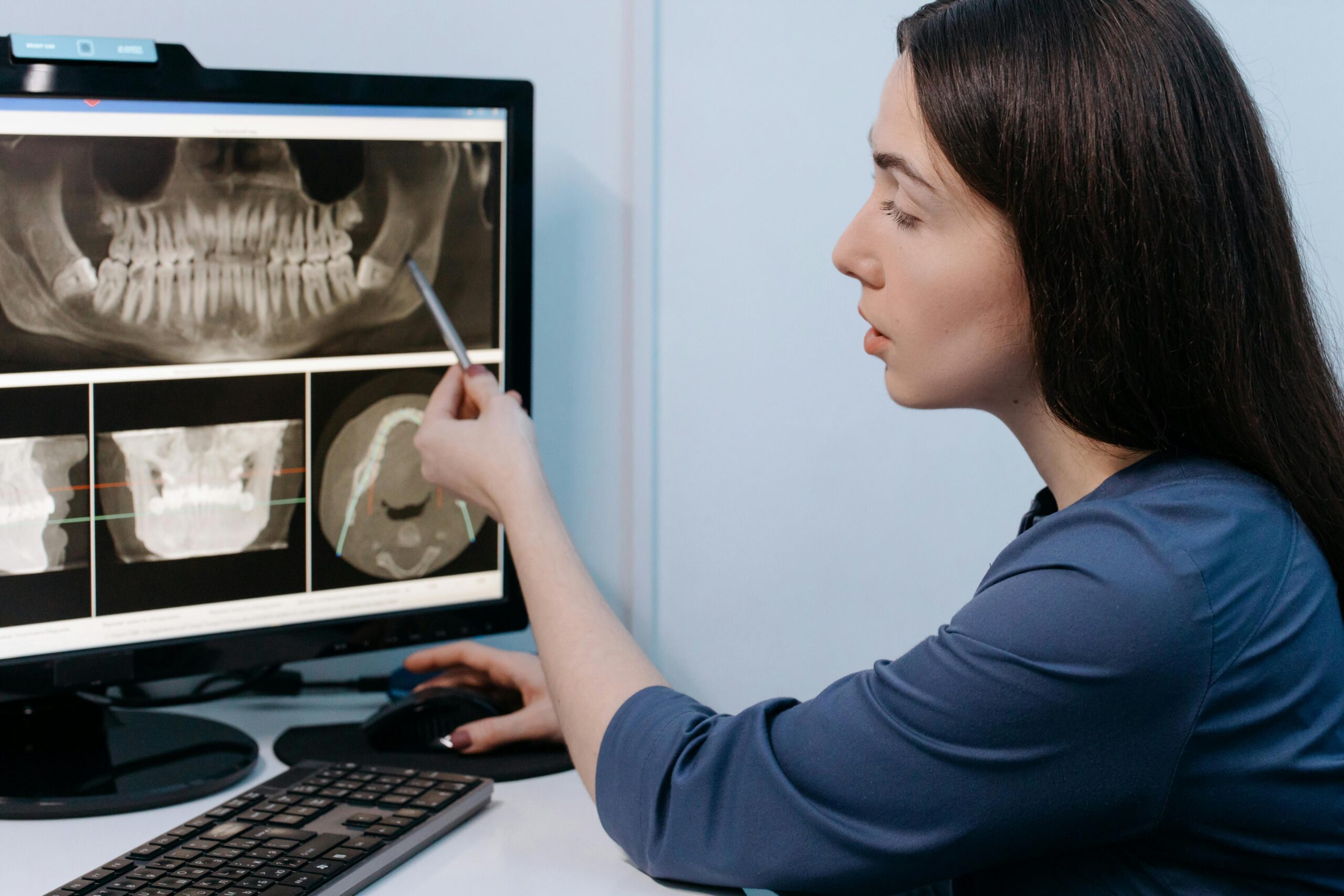
X-rays
A variety of techniques are used in diagnostic imaging, each suitable for a specific medical assessment. X-rays are one of the most common. They make it easier to identify infections or fractures by producing images of bones and particular tissues.
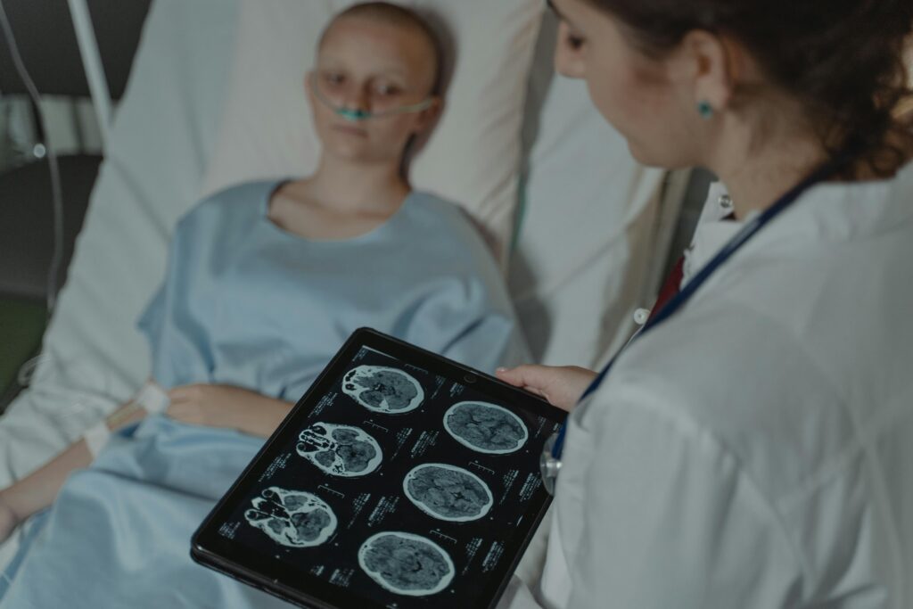
CT Scans
Compared to standard X-rays, CT scans offer a more detailed view because they combine multiple images into cross-sectional slices of the body. When diagnosing internal injuries or tumours, they are very helpful.
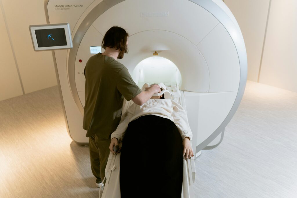
Magnetic Resonance Imaging (MRI)
The use of magnetic fields to produce a high-detail image of soft tissues makes MRI special. It is commonly used for brain, spinal cord, and joint examinations.
In nuclear medicine, tiny amounts of radioactive material are used to detect diseases at the cellular level, providing details that other imaging techniques might miss.
The Function and Significance of Diagnostic Images
Diagnostic images are crucial in modern medicine. By providing visual insights into the human body, they assist medical professionals in making precise diagnoses and treatment plans.
These images help diagnose a variety of conditions, such as fractures and tumours. They enable doctors to see inside structures without having to undergo invasive treatments. The patient’s risk is significantly reduced because it is non-invasive.
Early illness detection is also facilitated by diagnostic imaging. Early intervention can lead to better outcomes and higher recovery rates for patients.
These images are also necessary for medical professionals to interact with each other. They ensure that professionals collaborate effectively on complex cases, producing comprehensive treatment plans that are tailored to each patient.
In a world where time is often of the essence, diagnostic images speed up clinical decision-making. They are vital components of effective healthcare delivery systems worldwide, so their significance extends beyond appearance.
How to Decipher a Diagnostic Image
Reading a diagnostic image can be frightening at first, but it becomes more manageable with practice. First, become familiar with the common features of various imaging tests, including MRIs, CT scans, and X-rays.
Look for significant landmarks that indicate normal anatomy. It is simpler to identify abnormalities when one is aware of how healthy tissues look.
Observe the colour variations in the image. For example, shifting colours in MRI scans can reveal fluid levels or tissue density.
Note any abnormalities, such as lesions or lumps. These may point to potential health issues that need further research.
Consider using reference materials or software tools that can enhance image analysis. With time and practice, you will become an expert at interpreting diagnostic images.
Technological Developments in Diagnostic Imaging
The field of diagnostic imaging technologies is changing quickly. Innovations like 3D mammography and digital radiography are at the forefront. These advancements increase accuracy while reducing radiation exposure.
Artificial intelligence is revolutionising image analysis. Algorithms can now spot abnormalities faster than ever before, which raises the possibility of a diagnosis. This integration allows practitioners to focus more on patient care by optimising procedures in healthcare facilities.
The use of portable imaging devices is becoming more and more common. Handheld ultrasound devices reduce wait times and increase patient convenience by enabling doctors to perform examinations at the patient’s bedside.
Additionally, fusion imaging combines multiple modalities to offer a comprehensive understanding of medical conditions. For instance, by offering metabolic data in addition to anatomical details, PET-CT scans aid in precise treatment planning.
These technological developments not only improve diagnosis but also provide patients with quicker access to vital health status data. Future healthcare outcomes could be greatly enhanced by diagnostic imaging technology.
The Benefits and Drawbacks of Diagnostic Imaging
The benefits of diagnostic imaging are numerous. It provides important details about a patient’s health, revealing conditions that may not be visible with just a physical examination. Procedures like MRI and CT scans may save lives by detecting tumours, fractures, or internal bleeding early.
However, there are limitations to consider. Misunderstandings may arise from the use of diagnostic images if they are not properly analysed. False positives or negatives could result in missed diagnoses or unnecessary anxiety.
Cost is another factor to take into account; advanced imaging technology often comes with high expenses that insurance policies would not cover. Additionally, radiation exposure from certain procedures may eventually result in health issues.
Patients and medical professionals can choose better treatments if they are aware of the full range of diagnostic imaging, even though it is still evolving and improving medical procedures.
Final Thoughts
Diagnostic image comprehension is essential for anyone entering the medical field or simply seeking to increase their knowledge. These images are vital tools that support healthcare professionals in making educated decisions about diagnosis and treatment.
From MRIs to X-rays, each diagnostic image type provides a unique view of a patient’s health. Knowing the purpose of these images highlights how they enhance patient care and ensure accurate assessments.
Although it might be difficult, learning to read diagnostic images can be rewarding. People can acquire a sharp eye for detail via practice, which helps them comprehend and successfully communicate discoveries.
Check Also:https://coruzants.com/tech/ark-augmented-reality/